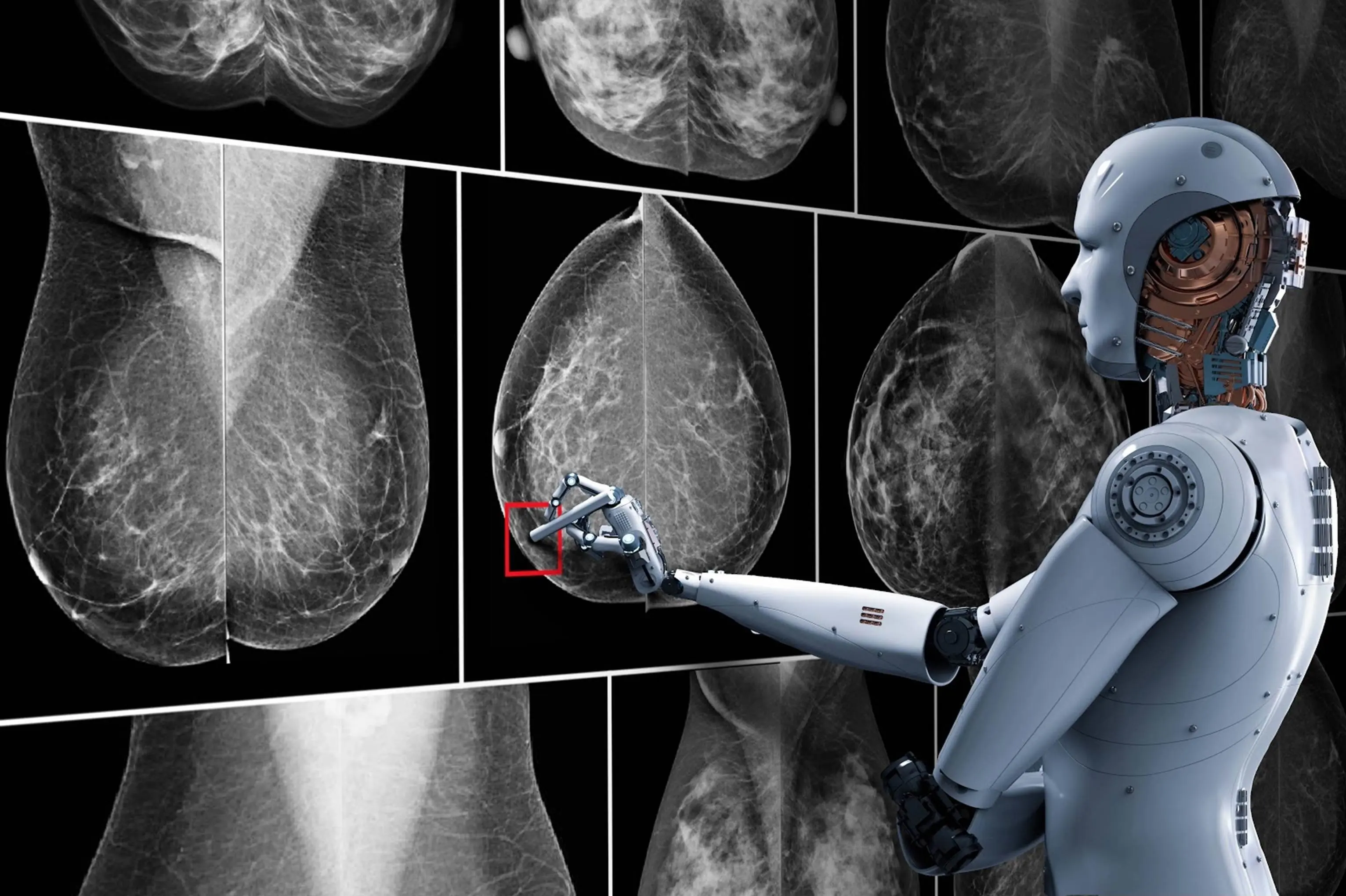
Today, screening for breast cancer, the most common cancer in women, is performed using mammography. Breast MRI and tomosynthesis are also frequently used in breast cancer screening, and the data obtained with these methods is difficult and time-consuming to evaluate due to their high-dimensional and complex nature. Numerous computer-aided auxiliary systems have been developed to increase radiologists' efficiency and accuracy in reading images.
In radiology, images are not simply images; they are actually the digital data that forms the basis of the image. With the emergence of deep learning algorithms in machine learning, computer vision, and medical image analysis, we are witnessing revolutionary developments. With the success of deep learning algorithms in image analysis, research on the subject has increased exponentially since 2012. The success of deep learning methods and convolutional neural network algorithms in image analysis in other fields has also begun to be used in medical image analysis and mammography reading.
One of the benefits of deep learning is the system's ability to self-learn; Instead of humans teaching computers to identify image features and perform related calculations, deep learning allows computers to learn image features themselves. In other words, deep learning methods have transitioned from teaching image features to computers learning image features themselves.
Computer-Aided Detection (CAD) software was developed in the early 1990s for breast cancer detection in mammography.
CAD systems are crucial for minimizing missed or misinterpreted lesions in mammography. New-generation deep learning-based CAD systems are contributing to the development of breast cancer screening programs. Research has shown that using AI-enhanced CAD as a decision support tool is more helpful to radiologists than the traditional approach. Furthermore, research has shown that breast radiologists achieve higher diagnostic performance with the help of AI-enhanced decision support systems compared to reading alone.
It is also important to understand the limitations of artificial intelligence systems. Machine learning systems, including deep learning systems, can only specialize in solving isolated tasks, while human intelligence makes decisions by synthesizing information from various sources and layers.
Artificial intelligence will undoubtedly impact radiology more rapidly than any other medical field. In just the last few years, various applications have been developed that match or even surpass human performance in certain image recognition tasks, such as breast imaging. The next step is for these applications to become part of daily routines once the necessary approvals and legal frameworks are completed.
In the future, deep learning-based AI systems will increase the efficiency and confidence levels of breast radiologists in their daily routines, freeing up time for radiologists to focus more on the patient's clinical experience. Radiologists will shift their focus from images to the patient with the help of decision support systems developed with AI applications.
At our clinic, we have been using Screenpoint's Transpara AI system, which received FDA approval in 2019 and whose development we contributed to by providing labeled data, for mammography and breast cancer screening since October 2018.

