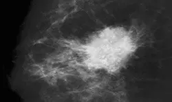


Galactography does not require any special preparation.
Approximately 18-25 milk ducts are opened per nipple. The duct where galactography is performed must be clear on the day of the examination to prevent medication being administered to the wrong duct. In other words, if there is no discharge, galactography is not appropriate. In that case, an X-ray of any of the ducts would be taken, but the goal is to see the inside of the duct causing the discharge in detail.
A mammogram is taken to reveal the inside of the duct by injecting a small amount of contrast material into the duct from the nipple where the discharge is occurring.
A filling defect (areas not filled with the medication) usually indicates a small mass. Most of these are intraductal papillomas. Some papillomas can develop into cancer and should be removed because they should be removed. Less than 10 percent of filling defects are caused by cancer. Galactography also aids surgical planning by identifying the location of the tumor within the duct.
It is important to have a recent mammogram of a patient undergoing galactography and compare it with it. It is also important to have normal mammograms performed without the use of medication, as the medication administered during galactography may prevent intracanal calcifications from appearing.

