Screening for breast cancer, the most common cancer in women, is performed with digital mammography.
In radiology, images are not simply images, but the underlying digital data. With the high success of deep learning algorithms in image analysis, the development of software for detecting breast cancer in mammograms has increased exponentially in recent years.
The fundamental advantage of deep learning over other machine learning systems is the system's ability to self-learn using labeled images. In other words, deep learning has enabled computers to learn image features themselves.
Computer-Aided Detection (CAD) software was developed in the early 1990s for breast cancer detection in mammography.
CAD systems are crucial for reducing the number of lesions missed or misinterpreted in mammography. CAD systems using new-generation deep learning are helping to increase the sensitivity of breast cancer screening programs. Research shows that breast radiologists achieve higher diagnostic performance with the help of decision support systems developed with artificial intelligence, compared to reading mammograms alone.
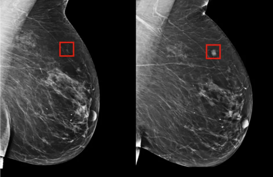
Artificial intelligence will undoubtedly impact radiology much more rapidly than other medical fields. In the last few years alone, numerous applications have been developed that match or even surpass human performance in certain image recognition tasks, such as breast imaging. The Transpara AI system, which we currently use in our clinic, acts as a secondary mammography reader, reading all mammograms and guiding the radiologist.
In the future, AI systems using deep learning will increase the efficiency of breast radiologists' daily diagnostic tasks, freeing up time for them, allowing them to focus more on the patient's clinical experience.
In our clinic, we have been using Screenpoint's Transpara AI system, which received FDA approval in 2019 and was developed by contributing labeled data, for breast cancer screening through mammography since October 2018.